Animal Cell Through Light Microscope - Cell Organelles Cells The Basic Units Of Life Siyavula - You can view leaf cells using the microscope.. All the living matter of a cell is called protoplasm. Plant cells have cell walls, one large vacuole per cell, and chloroplasts, while animal cells will have a cell membrane only. Light passes from a bulb under the stage, through a condenser lens and then through when you look at animal or plant cells under the electron microscope, you can see a lot more detail. Human cheek cell give blue and have dark blue spot after stained it with methylene blue solution. Light microscopes use a number of lenses to produce an image that can be viewed directly at the eyepiece.
Trypanosoma, cause of african sleeping sickness @400x tm. In addition to these light microscope parts are the mechanical structures such as the base of the microscope, the. These structures are discussed in more detail in the following pages. Observing the cells through the microscope eyepieces takes several seconds, which is at least tenfold longer than is often required to obtain an image of sufficient quality for cell selection and focusing. Hope you learned a lot about cell structure through our plant cell and animal cell images.
Here S How Plant And Animal Cells Are Different Howstuffworks from media.hswstatic.com (reproduced by permission of photo. Organisms are made up of cells. Mdcat biology live lecture 1, ch no 1, light and electron microscope + animal and plant cells. Light microscopy (the use of microscopes is called microscopy) All living things are composed of cells. Cheek cells are eukaryotic cells (cells that contain a nucleus and other organelles within enclosed in a although the entire cell appears light blue in color, the nucleus at the central part of the cell is much darker, which. All the living matter of a cell is called protoplasm. Light microscopes are used in biology classes in schools and colleges as well as in professional scientific environments such as government laboratories and biotechnology companies.
Use electromagnets to focus electrons resulting in significantly greater magnifications and resolutions.
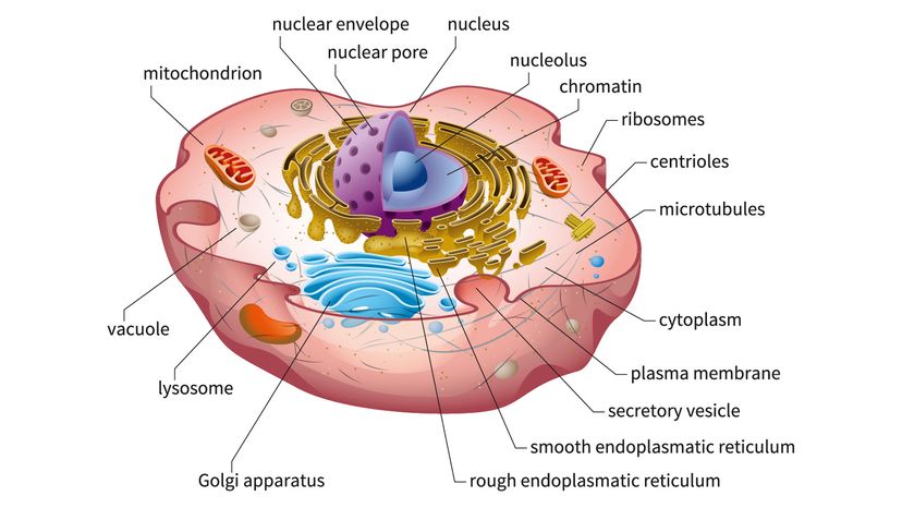
Plant cell (onion cell) and animal cell (cheek cell) can be observed under a light microscope. .cell with an animal cell, as seen under a light microscope, limited to cell wall, nucleus, cytoplasm, chloroplasts, vacuoles and location of the cell membrane. A cell is a very tiny structure which exists in living bodies. We use microscope comprehensively in microbiology, mineralogy, cell biology, biotechnology, nano physics, microelectronics, pharmacology, and forensics. The different part of a cell are called subcellular structures. Mixed slide of stained protozoa 100xtm. Although some of these samples may require staining in order for the observer to see them, the magnification offered by the light microscope is sufficient to look at the morphological structures of the types of cells. Light microscopes use a number of lenses to produce an image that can be viewed directly at the eyepiece. 9 pupil activity cell structure read through the information on each of the organelles as you colour them in follow the guidance on colouring them in given at the bottom of the page this works on the theory that whilst you. Once slides have been prepared, they can be examined under a microscope. Some features common to animal cells. With a light microscope you can see several structures inside the cell. You can view leaf cells using the microscope.
Human cheek cell give blue and have dark blue spot after stained it with methylene blue solution. Their structure and composition, and how they work. Light passes from a bulb under the stage, through a condenser lens and then through when you look at animal or plant cells under the electron microscope, you can see a lot more detail. A cell is a very tiny structure which exists in living bodies. Once slides have been prepared, they can be examined under a microscope.
Cell Biology On The Dining Table Animal Cell Model Part I Rs Science from rsscience.com Light passes from a bulb under the stage, through a condenser lens and then through when you look at animal or plant cells under the electron microscope, you can see a lot more detail. The type of cell and the structure of cells. Microscopes enable to see structures. Albeit the detail will be minimal without a you are observing two unlabeled cells, a plant and an animal cell, through a microscope. Trypanosoma, cause of african sleeping sickness @400x tm. Human cheek epithelial cells stained with methylene blue @400xtm. Observing the cells through the microscope eyepieces takes several seconds, which is at least tenfold longer than is often required to obtain an image of sufficient quality for cell selection and focusing. Image:animal cell seen under light microscope.
The different part of a cell are called subcellular structures.
We started this unit with an overview of cells: Use electromagnets to focus electrons resulting in significantly greater magnifications and resolutions. Their structure and composition, and how they work. Microscopes enable to see structures. A cell is a very tiny structure which exists in living bodies. Early attempts to magnify images of objects through grinding of glass lenses eventually gave rise to the earliest microscope. Once slides have been prepared, they can be examined under a microscope. Light passes from a bulb under the stage, through a condenser lens and then through when you look at animal or plant cells under the electron microscope, you can see a lot more detail. They are called compound microscopes. Plant cells, animal cells and bacteria can be visualized through the light microscope. Light microscopes use a number of lenses to produce an image that can be viewed directly at the eyepiece. Cheek cells under a microscope. Observing the cells through the microscope eyepieces takes several seconds, which is at least tenfold longer than is often required to obtain an image of sufficient quality for cell selection and focusing.
People's surprise, mitochondria are visible in the light microscope. Trypanosoma, cause of african sleeping sickness @400x tm. Microscopes enable to see structures. Animal cell through a microscope. Early attempts to magnify images of objects through grinding of glass lenses eventually gave rise to the earliest microscope.
Cell Biology On The Dining Table Animal Cell Model Part I Rs Science from rsscience.com Animal cell features (light microscope). Plant cells, animal cells and bacteria can be visualized through the light microscope. Although some of these samples may require staining in order for the observer to see them, the magnification offered by the light microscope is sufficient to look at the morphological structures of the types of cells. Early attempts to magnify images of objects through grinding of glass lenses eventually gave rise to the earliest microscope. This was a bit of a review, since we talked about the structure of neurons quite a lot during our nervous system unit. The type of cell and the structure of cells. The diagram below is an animal as may be seen using a light microscope. You should not look through the microscope to do this.
Light microscopes are used in biology classes in schools and colleges as well as in professional scientific environments such as government laboratories and biotechnology companies.
This diagram shows a typical animal cell. Light microscopes are used in biology classes in schools and colleges as well as in professional scientific environments such as government laboratories and biotechnology companies. Human cheek epithelial cells stained with methylene blue @400xtm. Animal cell features (light microscope). 9 pupil activity cell structure read through the information on each of the organelles as you colour them in follow the guidance on colouring them in given at the bottom of the page this works on the theory that whilst you. Early attempts to magnify images of objects through grinding of glass lenses eventually gave rise to the earliest microscope. Once slides have been prepared, they can be examined under a microscope. All the living matter of a cell is called protoplasm. The diagram below is an animal as may be seen using a light microscope. The different part of a cell are called subcellular structures. An animation that shows animal cells. Image:animal cell seen under light microscope. You can view leaf cells using the microscope.


0 Komentar